
Core Laboratories
Biomedical Research Imaging Center
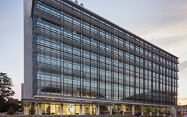
The HNG conducts its imaging at the Biomedical Research Imaging Center (BRIC) located in the new Marsico Hall at the UNC School of Medicine. Within the University of North Carolina, the BRIC houses state-of-the-art imaging equipment, directs innovative imaging research programs, and asserts outstanding skill and knowledge in imaging applications. Imaging hardware includes PET/MR,PET/CT, MRI, SPECT, Optical Imaging, and Ultrasound.
fMRI Data Analysis Core
Core Directors: Dr. Kathleen Gates and Dr. Joseph Hopfinger
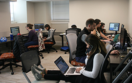
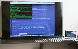
To enhance and support the existing needs of fMRI researchers and in anticipation of the expanding data processing needs of researchers doing human neuroscience studies, it is critical to have a core lab for data analyses. This common space enhances the sharing of expertise across human neuroimaging researchers within the Department of Psychology and Neuroscience, facilitating collaboration across labs and enhancing the training of students. An adjoining office for neuroimaging-focused post-doctoral fellows further builds cross-disciplinary interactions. Please contact Dr. Joseph Hopfinger or Dr. Kathleen Gates for more information.
Emotion Induction Core
Core Director: Dr. Kristen Lindquist
Many studies need to experimentally manipulate participants’ emotions by temporarily putting participants in a pleasant or unpleasant state and testing the effects on physiology or behavior. This common space enables the controlled manipulation of emotions using audiovisual stimuli (television and surround sound), social stimuli (a social “interview” set-up), and tactile stimuli (a “butt-kicker” that can cause vibration to participants’ chair). The room is outfitted with wireless and wired Mindware physiological recording hardware and software to non-invasively and continuously assess human cardiac, respiratory, blood pressure, facial electromyography and skin conductance responses. Eprime experimental software allows for the presentation of stimuli and collection of behavioral responses. A separate control room and data scoring space allow researchers to collect and monitor physiological data and to clean and score it. Please contact Dr. Kristen Lindquist for more information.
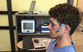
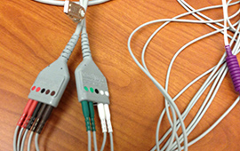
Electroencephalogram (EEG) Core
Core Directors: Dr. Margaret Sheridan and Dr. Joseph Hopfinger
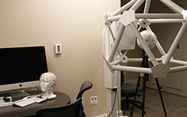
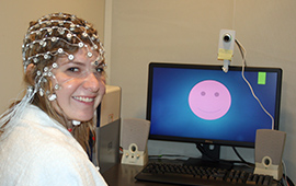
Recent innovations in data acquisition have made electroencephalogram (EEG) recording an ideal technology for investigating neural processes in young children and infants. This technology allows 128 electrodes to be applied to the scalp within a few minutes. The lab also houses a Geodesic Photogrammetry System that allows for precise localization of all 128 electrodes on the scalp simultaneously, enhancing neural source estimation of scalp recorded potentials, especially when used in combination with subject-specific MRI-derived head models. Electrical potentials recorded at the scalp reflect the summation of activity across neurons firing in unison generating local field potentials. Naturally occurring fluctuations in these signals can be measured in resting EEG and indexed in a time frequency analysis. Populations of children, such as those with and without ADHD, differ in their resting EEG patterns. In addition, perturbations in the EEG can be elicited and measured in experimental paradigms as event-related potential (ERP) waveforms. The EEG lab can be used in coordination with the neurostimulation facilities across the hall to perform transcranial alternating current stimulation (tACS), when the brain is stimulated at a specific EEG frequency. Please contact Dr. Joseph Hopfinger or Dr. Margaret Sheridan for more information.
Neurostimulation Core
Core Directors: Dr. Joseph Hopfinger and Dr. Charlotte Boettiger
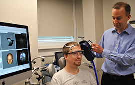
New methods of human neuroscience are allowing the testing of causal relations between brain and behavior by transiently stimulating regions of the brain. Transcranial magnetic stimulation (TMS) is a powerful technique that uses magnetic fields to stimulate precise regions of cortex. One variant of TMS can disrupt activity for only a few milliseconds, allowing one to assess not only the involvement of a brain region, but also the precise timing at which it is involved in that mental process. Another available method, transcranial direct current stimulation (tDCS) uses less powerful brain induction, essentially hyperpolarizing or hypopolarizing neuronal environments. This technique also provides critical information regarding the involvement of brain regions in cognitive processes. Finally, an exciting variant of the tDCS technique called transcranial alternating current stimulation (tACS) allows us to stimulate the brain at the specific frequencies that EEG studies have suggested to be involved in specific aspects of cognitive, perceptual, and social processing. Please contact Dr. Joseph Hopfinger or Dr. Charlotte Boettiger for more information.
Psychophysiology Core
Core Directors: Dr. Keely Muscatell and Dr. Michael Hallquist
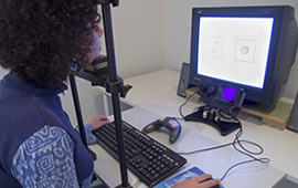
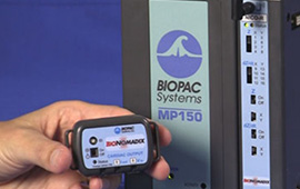
It is increasingly important to use multiple measures to assess attention and affect. This common suite of testing rooms enables the controlled manipulation and objective measurement of eye gaze, behavior, and peripheral physiology. Two separate testing rooms are outfitted with a S-R EyeLink system, and both wireless and wired psychophysiological hardware and software (including Biopac and Mindware systems). The mobile S-R system allows for the measurement of eye gaze fixation and saccades. Each testing room is additionally outfitted for the non-invasive and continuous assessment of human cardiac, respiratory, blood pressure, facial electromyography and skin conductance responses. Eprime experimental software allows for the presentation of stimuli and collection of behavioral responses. Noldus Observer software allows for the coding of behavioral responses. A separate control room allows researchers to collect and monitor physiological data. Please contact Dr. Kristen Lindquist for more information.